Foundational Discovery
1665
Hooke's Micrographia and the cell
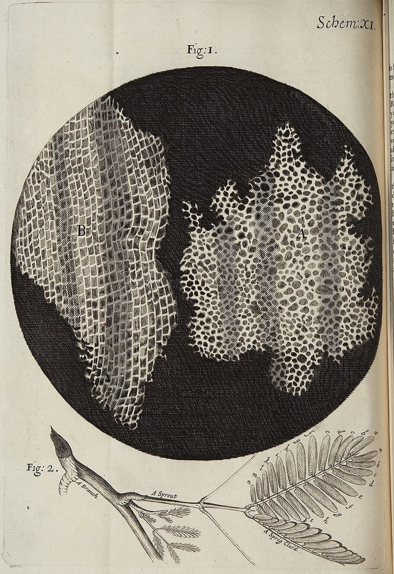
In Micrographia, published by the Royal Society in 1665, Robert Hooke described the box-like structure of cork under the microscope and called its compartments cells, the name later given to the basic unit of living things.
Key people
- Robert Hooke
- Curator of Experiments of the Royal Society; author of Micrographia
- Samuel Pepys
- Diarist who recorded reading Micrographia in January 1665
Source
Robert Hooke sharpened a penknife to a razor edge, cut an extremely thin slice from a clear piece of cork, and laid it on a black plate, since cork is white. With light cast on it through a deep plano-convex glass, it looked perforated like a honeycomb. The walls between the spaces were as thin, relative to the spaces, as the wax in a comb, and each long pore was divided by partitions into little boxes. Hooke called them pores or cells, and wrote in Observation 18 of Micrographia that they were the first microscopic pores he had ever seen, and perhaps the first anyone had seen.
He counted about 60 cells in an eighteenth of an inch, which gave him more than a million in a square inch and about 1,259,712,000 in a cubic inch. The structure explained cork's properties: it was light and floated, and it held air in a bottle, because its air was shut in separate boxes. He could find no passage from one cavity to the next, though he did not rule one out. The pith of elder and the stalks of fennel, carrots, burdock and ferns had a similar structure, and in green plants he saw the cells filled with juices.
The Council of the Royal Society ordered the book printed on 23 November 1664, and it appeared in 1665. Hooke was the Society's Curator of Experiments, and many of his observations answered requests made by Fellows at meetings over the previous year. He had been trained in drawing by the court painter Peter Lely and oversaw the 38 copperplate engravings, among them a flea and a louse, the louse on a plate that unfolds to nearly twice the size of the book. Another observation, on blue mould, gave the first published picture of a microorganism, the fungus Mucor, according to the Indiana University biologist Howard Gest.
Samuel Pepys sat up until two in the morning on 21 January 1665 reading it, and called it the most ingenious book he had ever read. Antoni van Leeuwenhoek, using single-lens microscopes of his own making, went on to describe protozoa and bacteria; Gest dates the two men's discovery of microscopic life to the years 1665 to 1683.
Keep exploring
 Read next · led to
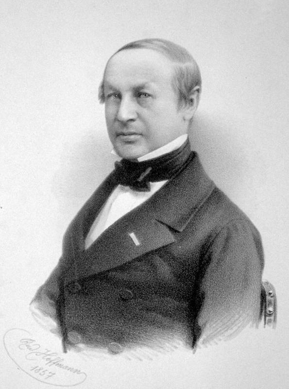
Schleiden and Schwann's cell theory (1839)
Hooke named the little boxes in cork cells, the name later given to the basic unit of living things. The cell theory story shows how Schleiden and Schwann made the cell the building block of all plants and animals.
Read this moment
Read next · led to
Schleiden and Schwann's cell theory (1839)
Hooke named the little boxes in cork cells, the name later given to the basic unit of living things. The cell theory story shows how Schleiden and Schwann made the cell the building block of all plants and animals.
Read this moment
 Today on The Clinical Times
Love to Dream recalls portable baby sound machine over fire and burn risk
Love to Dream has recalled its portable Sleep Machine because its lithium-ion battery can overheat while charging with an incompatible charger, creating a risk of fire and burns.
Read today’s top stories
Today on The Clinical Times
Love to Dream recalls portable baby sound machine over fire and burn risk
Love to Dream has recalled its portable Sleep Machine because its lithium-ion battery can overheat while charging with an incompatible charger, creating a risk of fire and burns.
Read today’s top stories
All 526 moments in the history of medicine. This one is in chapter 3, Anatomy and observation
Connected moments
How this moment links to others in the history of medicine, and what you will find in each.
-
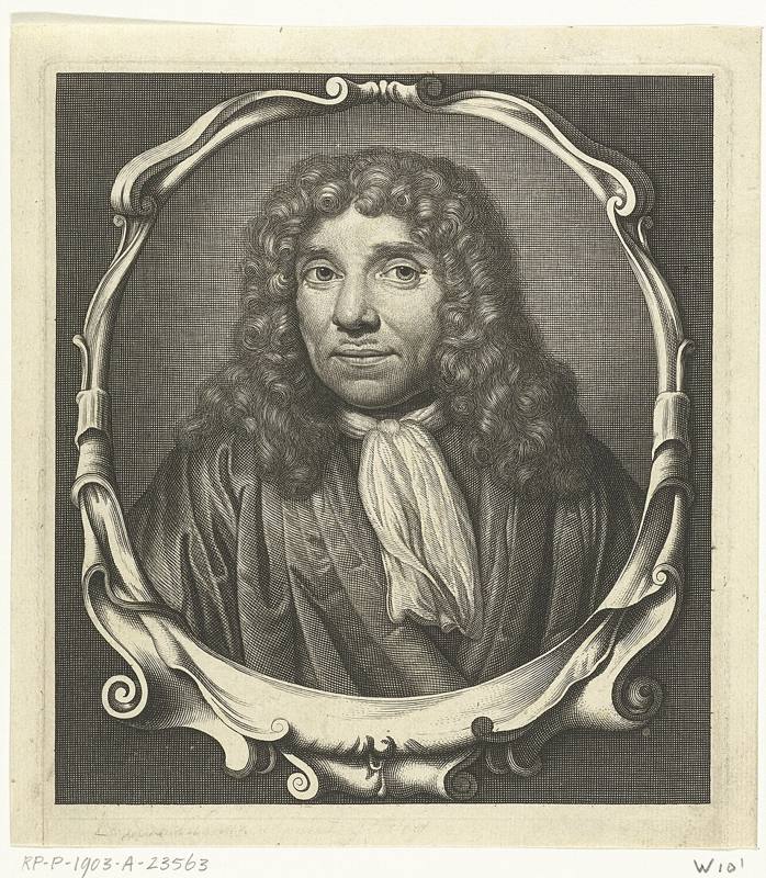 Same disease or problem1676Leeuwenhoek's AnimalculesHooke and Leeuwenhoek together opened up the microscopic world between 1665 and 1683. Leeuwenhoek's story shows his own single lens microscopes revealing protozoa and bacteria in water and in the plaque on his teeth.Read this moment
-
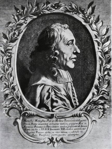 Same disease or problem1661Malpighi's Discovery of the CapillariesBoth used the new microscope to see structures too small for the eye, Hooke the cells of cork and Malpighi the capillaries of the lung. Malpighi's story shows how that finding completed the proof of the circulation.Read this moment