Infectious Disease
1949
Growing poliovirus in tissue culture
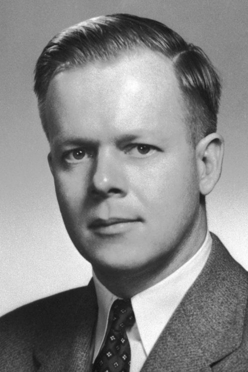
Enders, Weller and Robbins grew poliovirus in cultures of human tissue other than nerve cells, and the cell damage it caused showed under a microscope. Virus could now be grown and detected with far fewer monkeys, which made the polio vaccines possible.
Key people
- John F. Enders
- Directed the Children's Hospital laboratory in Boston where the work was done
- Thomas H. Weller
- Co-author of the 1949 paper; shared the 1954 Nobel Prize
- Frederick C. Robbins
- Co-author of the 1949 paper; shared the 1954 Nobel Prize
Source
In 1936 Albert Sabin and Peter Olitsky at the Rockefeller Institute tried to grow poliovirus in cultures of tissue from chick embryos, mice, monkeys and human embryos. Only human embryonic brain tissue kept the virus going, and the result was taken as proof that the virus lived in nerve tissue alone. Sabin and Olitsky did not want a vaccine made from brain tissue, fearing it could damage the nervous system of those who received it. Isolating and typing the virus from patients depended on injecting material into monkeys' brains, a slow and laborious method.
At the Children's Hospital in Boston, John Enders directed a research laboratory where Thomas Weller and Frederick Robbins worked with him after the Second World War. The group had already grown mumps and influenza viruses and had moved on to varicella, the chickenpox virus. They prepared flasks of human embryonic tissue, inoculated four with throat washings from chickenpox patients and, with the Lansing strain of poliovirus in a storage cabinet nearby, four more with poliovirus. The chickenpox virus did not grow. The poliovirus did.
Their report appeared in Science on 28 January 1949. They had added Lansing virus grown in mouse brain to flasks of embryonic skin and muscle from the limbs, which lacked intact nerve cells, and showed that it multiplied by injecting the fluids into mice; third-passage fluid paralyzed monkeys. It also grew in intestine and brain cultures, and two other strains followed. When the group later switched to roller tubes, a 1930s design by George Gey, the virus grew faster. In further work they saw that as the virus multiplied the cells changed and finally broke down, damage that was easy to see under a microscope, so live virus could be detected with much less need for monkeys. Immune serum blocked the growth, which made the cultures usable for antibody tests. Later the Boston group found that tissue removed at operations on children and adults worked as well, and they isolated virus directly from patient specimens.
Enders, Weller and Robbins shared the 1954 Nobel Prize in Physiology or Medicine for discovering that poliomyelitis viruses grow in cultures of various types of tissue. Presenting the prize, Professor S. Gard of the Royal Caroline Institute said human cell cultures let virologists study viruses that had no susceptible laboratory animal. In 1951 Jonas Salk and his colleagues at the University of Pittsburgh found that poliovirus could be grown on a large scale in monkey kidney cells, and Salk went on to make an injectable vaccine.
Keep exploring
 Read next · led to
Salk Inactivated Polio Vaccine Field Trial (Francis Report) (1955)
Growing poliovirus in tissue culture made the polio vaccines possible. The Salk story shows a trial of about 1.8 million children proving the killed virus vaccine worked.
Read this moment
Read next · led to
Salk Inactivated Polio Vaccine Field Trial (Francis Report) (1955)
Growing poliovirus in tissue culture made the polio vaccines possible. The Salk story shows a trial of about 1.8 million children proving the killed virus vaccine worked.
Read this moment
 Today on The Clinical Times
Love to Dream recalls portable baby sound machine over fire and burn risk
Love to Dream has recalled its portable Sleep Machine because its lithium-ion battery can overheat while charging with an incompatible charger, creating a risk of fire and burns.
Read today’s top stories
Today on The Clinical Times
Love to Dream recalls portable baby sound machine over fire and burn risk
Love to Dream has recalled its portable Sleep Machine because its lithium-ion battery can overheat while charging with an incompatible charger, creating a risk of fire and burns.
Read today’s top stories
All 526 moments in the history of medicine. This one is in chapter 5, Cures and codes
Connected moments
How this moment links to others in the history of medicine, and what you will find in each.
-
 Led to1960Sabin Oral Live-Attenuated Polio VaccineWith poliovirus grown in cell culture, Sabin could develop a live oral vaccine as well. His story shows the vaccine given by mouth that spread through mass campaigns.Read this moment
-
 Same person1963Edmonston-B measles vaccineJohn Enders went on to make a live measles vaccine from the Edmonston strain. That story shows American measles cases falling by more than 95 percent after 1963.Read this moment
-
 Same disease or problem1951The HeLa Cell LineThe Boston group grew poliovirus faster in George Gey's roller tubes, and Gey later grew the first endless human cell line from Henrietta Lacks's tumor. The HeLa story shows those cells taken without her knowledge.Read this moment